For Which of the Following Is Bitewing Film Used
Conventional radiography was used employing the following technical equipment. This type of Radiographic technique is used in recording the position and number of Supernumerary or Impacted teeth in.
Bitewing X Rays
Dear Bill Lets start with the bottom line.
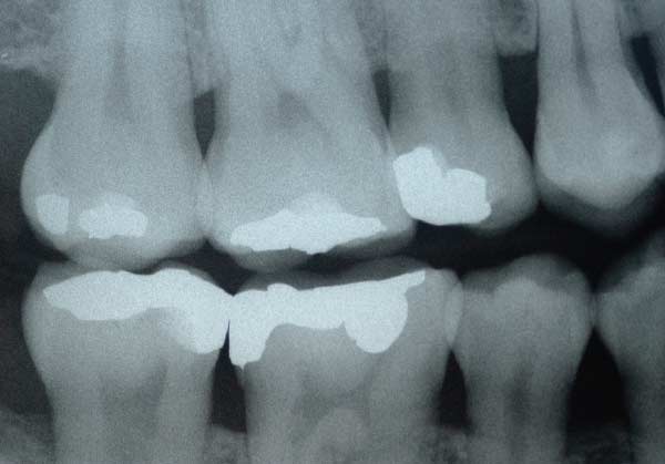
. Girardelli X-30 for the developer. The bitewing view is taken to visualize the crowns of the posterior teeth and the height of the alveolar bone in relation to the cementoenamel junctions which are the demarcation lines on the teeth which separate tooth crown from tooth root. This film shows the crowns of both the maxillary and mandibular teeth and a portion of the roots.
9State the basic rules for the bitewing technique. Scope1 that has an encryption type of Microsoft-managed keys. Which of the following is the correct vertical angulation used for the bitewing technique.
The ___ is the coronal portion of alveolar bone found between teeth. -Receptor placed to cover the area of the tooth to be examined receptor placed parallel to crowns vertical angulation directed at 10 degree horizontal angulation must be directed through contact area of teeth x-ray beam must be centered on the receptor. Bitewing radiographs were taken at the time of the clinical examination.
Terms in this set 18 The bitewing view is used for detecting. A periapical view shows the tooth from the occlusal surface or incisal edge to the. An object is ___ if its moving or lying in the same plane is always separated by the same distance.
A form of entertainment information etc composed of such a sequence of images and shown in a cinema etc. The following information is printed on the back. 31-35 show the evolution of dental X-ray film throughout the years as described by Campbell 70 and other reviews.
Carestream Insight IP-21 for the film. As the name suggests this films is used to record the Maxilla or Mandible from the occlusal surface showing all the teeth Oclussaly. The object tooth should be as far as practical from the target.
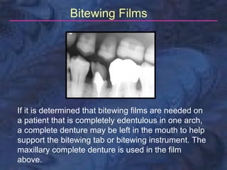
The bitewing receptor is used. A premolar film and molar film are normally taken on each side for a total of four posterior bitewing films. And Carestream Readymatic for the developer and fixer.
An intraoral full-mouth survey FMX on an adult consist of --------- images. Routine bitewing radiographs are commonly used to examine for interdental caries and recurrent caries under. A sequence of images of moving objects photographed by a camera and providing the optical illusion of continuous movement when projected onto a screen.
Use the EP size 2 film to take bitewing views. Bblue Rinn type holder is used to stabilize the film sensor in the patients mouth. Periapical Examination- is used to examine the entire tooth crown and root and supporting bone.
Interproximal decay and periodontal disease. While the levels of radiation associated with bitewing digital x-rays are low dentists still take precautions to limit patients exposure by only taking x-rays when needed and by. In which position should the patient be placed infor bitewing radiographs.
Interproximal Examination- is used to examine the crowns of both maxillary and mandibular teeth on a single image. The dental specialists who routinely use exraoral radiographs are. ___ means cutting across or through.
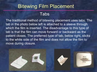
These are sticky paper tabs that fold and stick to the film whilst giving a horizontal surface for the patient to bite on. Radiation exposure using bitewing digital x-rays is minimal often quoted as being 80 percent less than conventional methods of dental x-rays involving film. You create the following encryption scopes for storage1.
If the beam missed part of the film the result is. Bitewings are used to detecthint- there are 3 things Calculas caries and faulty restorations. To present a complete image in the.
Bone about 2 to 3 mm beyond the apex. There are four sizes of Bitewing radiographic films based on the position and the age of the patient it is used in. Evidence-based research clearly shows that used judiciously bitewing x-ray imaging is the method of choice for determining the presence and extent of decay in the areas where back.
Occlusal film E 0 IO 41 or 42 DF 50 This film is considerably larger than the periapical film. Contact areas- where adjacent tooth surfaces meet. Often much comfier for the patient but ensure that the film is positioned well before the patient bites else if they hit the occlusal surfaces of the upper molars first instead of the palate they tend end up biting on the film flat.
The film typically used for the intraoral bitewing exam falls into three film speed classes - D slowest E and F-speed fastest. In this study in order to separate each tooth in the bitewing image first horizontal projection was applied to separate the upper and lower rows of teeth in the bitewing film into two photos while vertical projection was applied to separate the individual teeth from the upper and lower rows of teeth into a single individual tooth photo. Oral surgeons and Orthodontists.
The following timeline Table 1 and figures Fig. As modifier. Radiographic x-ray imaging is an integral tool in the assessment and diagnosis of dental caries tooth decay and periodontal gum disease.
- manufacturers name film speed the number of films in the packet one or. 13 - Which of the following intensifying screen must be used with a green - sensitive film. An area of a tooth that touches an adjacent tooth in the same arch is the ___.
The bitewing film is used for the detection of interproximal caries and the condition of the alveolar bone. List the 2 ways a film can be stabilized in the bitewing technique and identify which one is recommended for bitewing exposures. - Receptor placed to cover the area.
The types of film used by dental practices in this survey.
Radiology Bitewing Technique

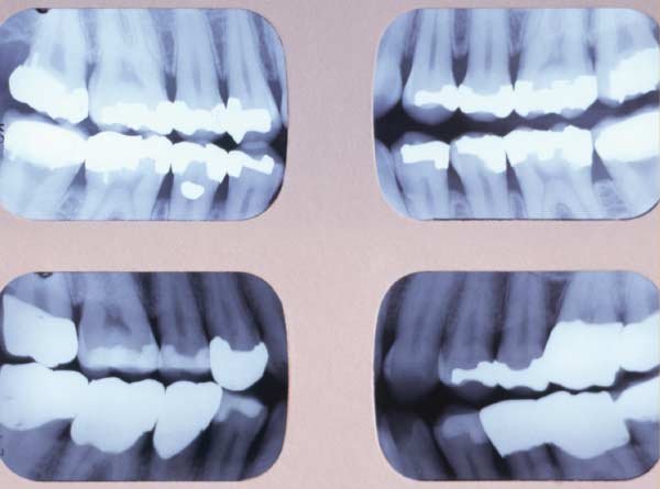
Examples Of Bitewing Films With The Correct Number Of Teeth Download Scientific Diagram
Bitewing X Rays
Radiology Bitewing Technique

Comments
Post a Comment